In Vivo Biocompatibility Imaging

Overview
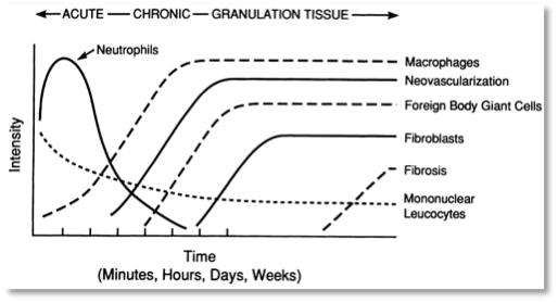
The foreign body response, instigated by monocytes and neutrophils, is initiated when a biomaterial is implanted, followed by propagation of fibroblasts and vascular endothelial cells.[1] Several complications can arise from the infiltration of inflammatory cells, such as: bio-instability of glucose sensors;[2] overgrowth of encapsulated pancreatic islets for diabetes therapy causing ischemia and, eventually, necrosis of the islets;[3] and constrictive fibrosis of implants.[4] This process is summarized in figure 1. Granulation tissue, fibrous connective tissue that replaces a fibrin clot in healing wounds consisting of Type III collagen, is created and modified by fibroblasts and may appear as early as 3 to 5 days surrounding the implant.[1] Molecular level understanding of this process in vivo and kinetically is poorly understood. This project aims to address the lack of insight into these processes through in vivo imaging of the foreign body response.
Approaches
Biocompatibility of polymers has been interrogated through in vivo fluorescence imaging.[6] However, molecular level quantification has yet to be described. Through the use of complementary imaging techniques – sum frequency generation (SFG), second harmonic generation (SHG) and two-photon excited fluorescence (TPEF) – the dynamic processes of the foreign body response will be elucidated.
SFG and SHG are non-linear optical processes in which two beams are overlapped temporally and spatially on a surface whereupon a third beam is generated at the sum frequency of the incoming two beams. For SHG, the frequencies of the two incoming beams are identical while in SFG, the frequencies are different. Media with centrosymmetry do not exhibit SFG or SHG under the electric dipole approximation. Two of the main advantages of SFG and SHG are that there is no photobleaching since the incoming light is not in resonance with the molecule and that labeling of the molecule is not required, unlike with fluorescence imaging.
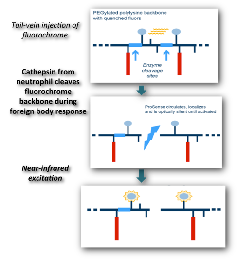
Neutrophil and macrophage activation can be imaged through protease–activatable fluorescence sensors, commercially known as Prosense-680. Prosense-680 is a protease-activatable fluorescence sensor based on a polymeric scaffold that allows imaging of cathepsin B activity (and to a lesser extent cathepsins K, L, and S), allowing for detection of neutrophils and macrophages. The fully assembled Prosense-680 scaffold consists of near infrared fluorochromes, specific lys-lys peptide substrates and partially methoxypegylated graft copolymers. Proteolytic cleavage of the scaffold releases the fluorochromes and results in extensive fluorescence generation (dequenching) at 700 nm. The major advantage of this technique is its ability to monitor multiple time points. Kinetic studies monitoring the changes in macrophage and neutrophil activation will yield insight into what material properties intensify phagocytosis.
Current Students
Anuraag Boddupalli, Materials Science Ph.D. student
Figure 1. The temporal variation in the acute inflammatory response, chronic inflammatory response, granulation tissue development, and foreign body reaction to implanted biodegradable microspheres.[5]
References
-
1.Kindt, Goldsby, Osborne Kuby Immunology. New York: W. H. Freeman and Company, 2007
-
2.P. H. Kvist, T. Iburg, M. Bielecki, M. Gerstenberg, T. Buch-Rasmussen, E. Hasselager, H. E. Jensen. Biocompatibility of electrochemical glucose sensors implanted in the subcutis of pigs. Diabetes Technology & Therapeutics, 2006, 8, 463.
-
3.P. De Vos, B. J. Haan, R. Van Schilfgaarde. Factors causing failure of islets in nonovergrown capsules. Transplantation Proceedings, 1998, 30, 496.
-
4.P. Wilflingseder, A. Propst, G. Mikuz. Constrictive fibrosis following silicone implants in mammary augmentation. European Journal of Plastic Surgery, 1974, 2, 215.
-
5.J. M. Anderson, M. S. Shive. Biodegradation and biocompatibility of PLA and PLGA microspheres. Advanced Drug Delivery Reviews, 1997, 28: 5.
-
6.K. M. Bratlie, T. T. Dang, S. Lyle, M. Nahrendorf, R. Weissleder, R. Langer, D. G. Anderson. Rapid Biocompatibility Analysis of Materials via In Vivo Fluorescence Imaging of Mouse Models, PLoS ONE 2010, 5, e10032.
Figure 2. Protease activatable probe for whole body fluorescence imaging. The probe has a poly-L-lysine backbone that has been PEGylated to increase circulation time. Cy5.5 fluorophores are conjugated on the lysine side chains, which are in close proximity so that the probe self-quenches in the intact state. Upon exposure to cathepsins - released by leukocytes - the poly-L-lysine backbone is cleaved and a fluorescent signal can be measured.[6]
The Bratlie Research Group
Department of Materials Science and Engineering
Department of Chemical and Biological Engineering
Iowa State University, Ames, IA 50011
© 2011 Kaitlin Bratlie