Macrophage Reprogramming

Current Students
Jenny Li, Materials Science & Engineering Ph.D. student
References
-
1.H.C. Bygd, K.D. Forsmark, K.M. Bratlie The significance of macrophage phenotype in cancer and biomaterials, 2014, Clinical and Translational Medicine 3, 62.
-
2.M. C. Schmid, J. A. Varner. Myeloid cells in the tumor microenvironment: Modulation of tumor angiogenesis and tumor inflammation. Journal of Oncology, 2010, 2010, 201026.
-
3.S. Donnelly, S. M. O. Neill, M. Sekiya, G. Mulcahy, J. P. Dalton. Thioredoxin peroxidase secreted by Fasciola hepatica induces the alternative activation of macrophages. Infection and Immunity, 2005, 73, 166.
-
4.S. Gordon, Alternative activation of macrophages. Nature Reviews Immunology, 2003, 3, 23.
-
5.M. G. Nair, D. W. Cochrane, J. E. Allen. Macrophages in chronic type 2 inflammation have a novel phenotype characterized by the abundant expression of Ym1 and Fizz1 that can be partly replicated in vitro. Immunology Letters, 2003, 85, 173.
-
6.A. Mantovani, S. Sozzani, M. Locati, P. Allavena, A. Sica, Macrophage polarization: tumor-associated macrophages as a paradigm for polarized M2 mononuclear phagocytes. Trends in Immunology, 2002, 23, 549.
-
7.R. D. Stout, S. K. Watkins, J. Suttles. Functional plasticity of macrophages: in situ reprogramming of tumor associated macrophages. Journal of Leukocyte Biology, 2009, 86, 1105.
-
8.J. Shapiro et al. International Trial of the Edmonton Protocol for Islet Transplantation. The New England Journal of Medicine, 2006, 335, 1318–1330.
-
9.K. M. Bratlie, R. L. York, M. A. Invernale, R. Langer, D. G. Anderson. Materials for Diabetes Therapeutics. Advanced Healthcare Materials, 2012,1, 267–284.
-
10.D. Akilbekova, R. Philiph, A. Graham, K. M. Bratlie Macrophage reprogramming: Influence of latex beads with various functional groups on macrophage phenotype and phagocytic uptake in vitro, 2015, Journal of Biomedical Materials Research Part A 103A, 262.
-
11.D. Wang, N. Phan, C. Isely, L. Bruene, K.M. Bratlie Effect of surface modification and macrophage phenotype on particle internalization, 2014, Biomacromolecules 15, 4102.
-
12.D. Wang, K.M. Bratlie On the influence of polymer chemistry on cytokine secretion from polarized macrophages, 2015, ACS Biomaterials Science & Engineering accepted
Overview
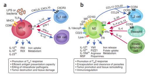
Two pathways for activation of macrophages exist.[1] One of these pathways is through interferon-γ (INF-γ) or lipopolysaccharide (LPS)[2], which is known as the classically activated M1 macrophage pathway. M1 macrophages are known as pro-inflammatory cells.[3]The alternative activated M2 macrophages are generated upon exposure to interleukin-4 (IL-4).[1,4] Tumor-associated macrophages (TAMs) are M2 polarized cells and promote tumor growth through the release of angiogenic - blood vessel forming - molecules.[5] This polarization is illustrated in Figure 1.
In vitro studies have shown that Th 2 macrophages can be reprogrammed to promote tumor regression through delivery of cytokines and chemokines. IL-12 has successfully reversed the polarization of TAMs in culture.[6]
Another application is in producing therapeutics for type 1 diabetes. Type 1 diabetes is an autoimmune disease in which the host’s immune system destroys insulin-producing pancreatic islets. This disease can be reversed through transplantation of insulin producing cells. As with any such transplant, an immune response is invoked which requires long-term immunosuppressive drugs to protect the transplanted tissue.[7] Alternatively, the islets can be encapsulated in a semi-permeable container, isolating and protecting them from the immune system. This is diagramed in Figure 2. The longevity of encapsulated cells largely depends on two factors: (1) availability of nutrients from nearby blood vessels or capillaries and (2) the amount of cytotoxic cytokines and chemokines secreted by immune cells.[8] Polarizing macrophages to alternatively activated macrophages, which have an increased expression of pro-angiogenic molecules and a decreased expression of pro-inflammatory molecules, presents an appealing method by which to combat both of these problems.
Approaches
We engineer polymers that induce either Th 1 or Th 2 polarization of macrophages for drug delivery to TAMs or angiogenesis for artificial organs.[9] The molecular responses of cells to these polymers is optimized in vitro and the tissue response is monitored through in vivo imaging methods to visualize phagocytic responses [10, 11, 12].
Figure 1. Schematic illustrating two of the possible polarization pathways of macrophages. (a) Classically activated M1 macrophages arise as a response to interferon-γ (INF-γ) or lipopolysaccharide (LPS). M1 cells produce pro-inflammatory cytokines including tumor necrosis factor-α (TNF-α), IL-12, reactive nitrogen intermediates (RNI), reactive oxygen intermediates (ROI), and several others listed in the figure. These macrophages are part of the polarized type I T helper (Th1) response. (b) Alternatively activated M2 macrophages are generated in response to IL-4, IL-13, IL-10, and glucocorticoids. Pro-angiogenic M2 cells release IL-1β, IL-6, IL-8, vascular endothelial growth factor (VEGF), and matrix metalloproteinases. These cells are part of the polarized type II T helper (Th2) response. FR, folate receptor; GR, galactose receptor; IFN-γR, IFN-γ receptor; IL-1decoyR, IL-1 decoy receptor; MHCII, major histocompatibility complex class II; MR, mannose receptor; SR, scavenging receptor; ST2, receptor; CXCL, chemokine (C-X-C motif) ligand; NK cell, natural killer cell; TLR, toll like receptor; CD, cluster of differentiation; CCL, chemokine (C-C) motif ligand; LYVE-1, lymphatic vessel endothelial hyaluronan receptor; YM1 and FIZZ1 are gene markers for M2 macrophages.[4]
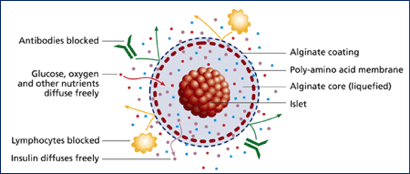
Figure 2. Schematic of encapsulated islets for type 1 diabetes therapy. The capsule allows nutrients, waste, and insulin to pass freely while protecting the islets from recognition by immune cells.
The Bratlie Research Group
Department of Materials Science and Engineering
Department of Chemical and Biological Engineering
Iowa State University, Ames, IA 50011
© 2011 Kaitlin Bratlie